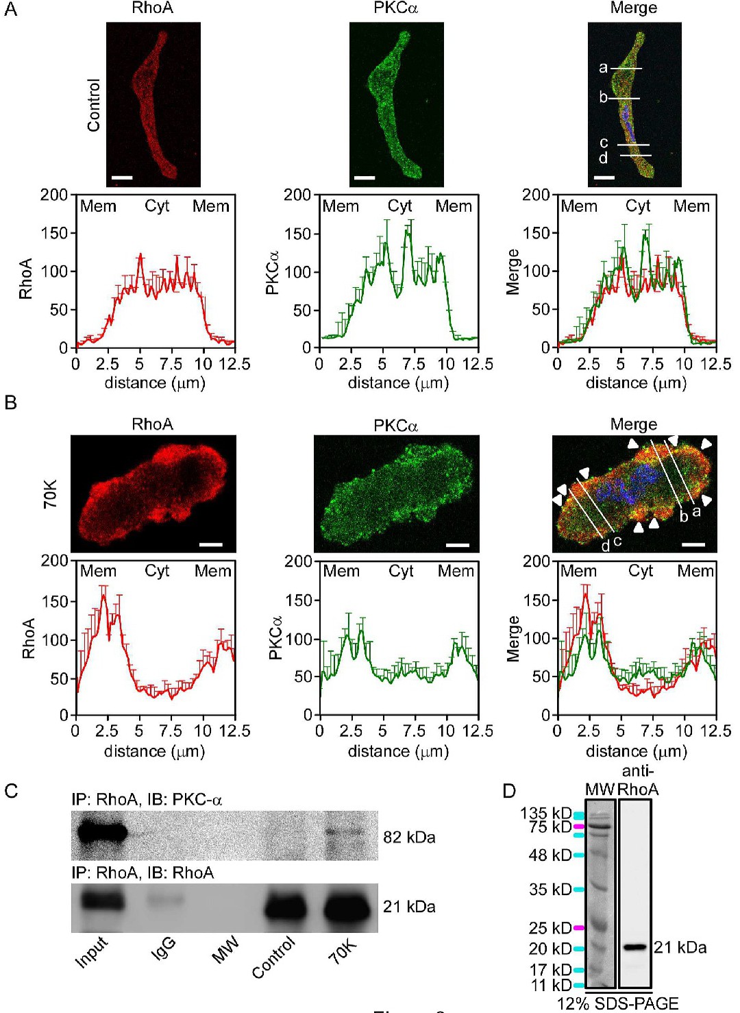
Fig. 3. Depolarization-induced RhoA and PKCα translocation to the periphery of rat arteries and coimmunoprecipitation of both proteins. Confocal immunofluorescence images (top panels) and immunofluorescence analyses (bottom panels) illustrating PKCα and RhoA distribution (PKCα, green; RhoA, red; cell nucleus, blue) in resting conditions (control, A) or 70K (B) in VSMCs from femoral arteries. Analyses were performed across four lines in different parts of the cell (a, b, c and d in merged images). Scale bars represent 8 µm (A) or 4 µm (B), (n = 3). Data represent mean ± S.E.M. (C) Effect of depolarization on PKCα-RhoA interaction from dispersed cells of femoral and aortic arterial tissue. Representative experiment that illustrates association of RhoA with PKCα (n = 3). Immunoprecipitation was performed with an anti-RhoA antibody, and western blots using PKCα (top) or RhoA (bottom) antibodies. (D) Western blot confirming that the antibody detects a band of the expected molecular weight (~ 21 kDa). Molecular weight standards (visible, left columns) for 12% SDS-PAGE and western blotting (chemiluminescence, right columns) of rat arterial homogenates (n = 3).